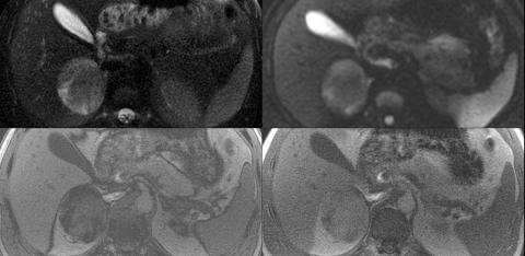
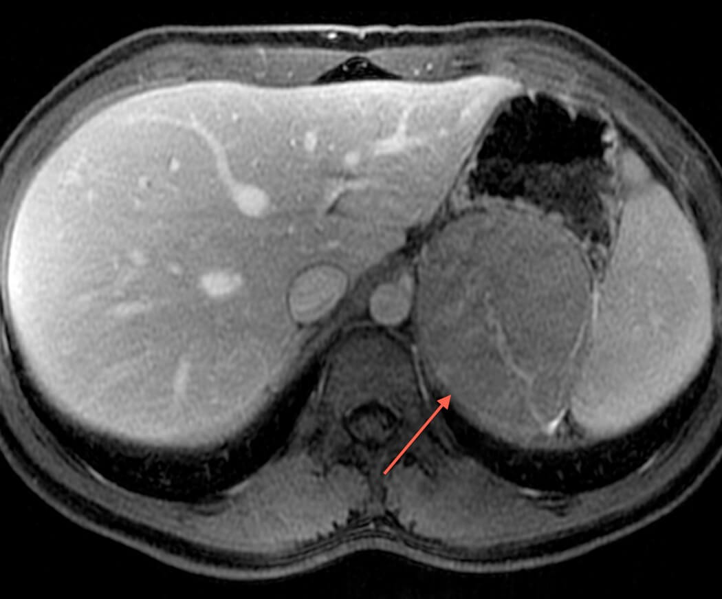
MRI is valuable when there is a clinical concern for an adrenal mass. This is often in the context of potential secondary hypertension. MRI provides unparalleled soft-tissue resolution and in a single scan can evaluate renal arteries for narrowings that cause hypertension and detect the presence of a renal or adrenal mass. Further, it can detect the presence of lipids in adrenal adenomas and myelolipomas to distinguish these lesions from metastases and pheochromocytomas. For renal lesions, the soft-tissue characterization of MRI can classify lesions as benign or malignant, and further, for malignant lesions can subtype the lesions.
References
Marin D, Dale BM, Bashir MR, et al. Effectiveness of a three-dimensional dual gradient echo two-point Dixon technique for the characterization of adrenal lesions at 3 Tesla. Eur Radiol 2012; 22:259-268. PubMed link
Schindera ST, Soher BJ, Delong DM, Dale BM, Merkle EM. Effect of echo time pair selection on quantitative analysis for adrenal tumor characterization with in-phase and opposed-phase MR imaging: initial experience. Radiology 2008; 248:140-147. PubMed link