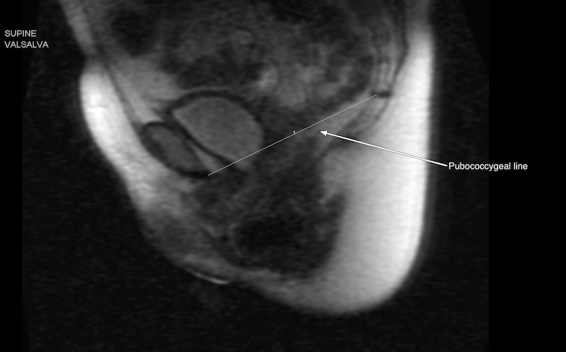
Sagittal MR image obtained of a supine patient performing the Valsalva maneuver. The descent of the urinary bladder, vagina, and rectum are compared to the pubococcygeal line.
An enterocele seen by dynamic MRI, and another case showing a posterior perineal hernia.
References
Reiner CS, Tutuian R, Solopova AE, Pohl D, Marincek B, Weishaupt D. MR defecography in patients with dyssynergic defecation: spectrum of imaging findings and diagnostic value. Br J Radiol; 84:136-144. PubMed link
Colaiacomo MC, Masselli G, Polettini E, et al. Dynamic MR imaging of the pelvic floor: a pictorial review. Radiographics 2009; 29:e35. PubMed link
Hecht EM, Lee VS, Tanpitukpongse TP, et al. MRI of pelvic floor dysfunction: dynamic true fast imaging with steady-state precession versus HASTE. AJR Am J Roentgenol 2008; 191:352-358. PubMed link
El Sayed RF, El Mashed S, Farag A, Morsy MM, Abdel Azim MS. Pelvic floor dysfunction: assessment with combined analysis of static and dynamic MR imaging findings. Radiology 2008; 248:518-530. PubMed link
Seynaeve R, Billiet I, Vossaert P, Verleyen P, Steegmans A. MR imaging of the pelvic floor. JBR-BTR 2006; 89:182-189. PubMed link
Hetzer FH, Andreisek G, Tsagari C, Sahrbacher U, Weishaupt D. MR defecography in patients with fecal incontinence: imaging findings and their effect on surgical management. Radiology 2006; 240:449-457. PubMed link
Birchard KR, Fielding JR. Fecal incontinence in the postreproductive woman. Curr Womens Health Rep 2003; 3:405-409. PubMed link